How Sagittal Section of Skull Models Are Reconstructed From Digital Anatomical Data
The clean cut surface of a printed skull conceals a long digital workflow. Sectional source images must be interpreted as anatomy, converted into continuous geometry, divided at the intended plane, prepared with visible surface information, and manufactured without losing the relationships established earlier. Reconstruction is therefore a chain of dependent decisions. Accuracy at the final print begins with the organization of the source data and continues through every transformation applied to it.
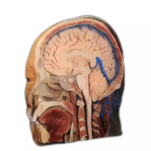
Defining the Source and the Sagittal Plane
High-precision digital human datasets, organized as sectional information, supply the starting material for reconstructing sagittal section of skull. Each source image records one level, while the complete skull requires corresponding boundaries to be followed through the sequence.
The intended cut plane must also be defined within the reconstructed coordinate system. A mid-sagittal section passes through the median region from front to back, but the digital model still needs consistent orientation before that division is applied. Small changes in plane position may alter which internal structures appear on the surface.
Source quality affects the boundaries available for reconstruction. Closely sampled, clearly differentiated anatomy provides more information for following thin plates, curved walls, and openings. The dataset remains an anatomical record rather than a finished printable object.
Before segmentation begins, orientation and sequence order require verification. The sectional data must be organized consistently so that anatomical boundaries remain spatially aligned during reconstruction. A consistent coordinate framework keeps the later geometric skull aligned with the anatomical planes represented in the source.
Converting Sectional Voxels Into Geometry
Segmentation identifies the skull within consecutive images and separates it from neighboring anatomy. Those labeled regions supply the basis for building a geometric surface. Continuity must be maintained where a structure changes outline, divides, narrows, or disappears from one level to the next.
The resulting mesh describes the internal and external surfaces in three dimensions. Irregularities caused by imaging or segmentation may require refinement, but smoothing cannot be allowed to close a small opening or erase a sharp boundary that carries anatomical meaning.
Geometry must also be printable. Very thin components, deep recesses, or isolated fragments may need careful surface preparation and support planning. Manufacturing requirements enter at this stage without changing the intended relationship between the cranial exterior and the sagittal interior.
The section operation must leave a closed, interpretable surface rather than an open digital shell. Edge cleanup and surface continuity are checked around the cut so that the printed plane represents anatomy clearly and does not expose accidental gaps created by the mesh.
Generating Texture Maps From Surface Voxels
For a sagittal section of skull, texture preparation links the reconstructed surface back to voxel information retained in the sectional source. DIGIHUMAN uses the resulting map to place visible source information at the corresponding geometric location.
Texture does not repair incorrect anatomy. If a boundary is misplaced in the geometry, a realistic surface pattern will only cover the error. Geometric review and texture review therefore address separate risks before they are combined.
The cut surface and the external skull may also require different visual treatment. The section needs clear internal boundaries, while the exterior must retain recognizable landmarks. Texture mapping can preserve that distinction without detaching the two surfaces from their shared coordinate system.
Registration is checked by comparing recognizable points on the texture with their geometric locations. A visible feature that drifts across a ridge or opening signals a mismatch between the surface map and the mesh, even when both files appear plausible when reviewed separately.
Full-Color, Multi-Material Fabrication
DIGIHUMAN uses full-color and multi-material technology in its broader anatomical printing process. After the geometry and surface information have been prepared, DIGIHUMAN uses full-color, multi-material 3D printing with inkjet and light-curing technologies to produce the physical model.
Color can distinguish neighboring regions or maintain visible information derived from the source. Multiple materials can assign different hardness, transparency, or opacity where the model design calls for those effects. These options remain subordinate to the anatomy established in the data.
Printing introduces its own controls, including alignment between layers, support for narrow structures, curing, and surface finish. A successful fabrication keeps the planned sagittal plane readable while preserving the external form used for orientation.
Post-print inspection returns to the anatomical questions established at the beginning. The cut plane is examined for continuous boundaries, the exterior for recognizable landmarks, and the whole object for distortion or material artifacts that could change the apparent spatial relationship.
Preserving Detail for Repeated Observation
By the time the sagittal section of skull is ready for use, several stages have been combined: dataset selection, anatomical segmentation, geometric reconstruction, definition of the section plane, surface-voxel extraction, texture mapping, and full-color multi-material printing. Each stage contributes a different part of the final evidence.
The mid-sagittal skull in the DIGIHUMAN anatomical-model range is intended for repeated observation, comparison, and teaching. Its physical stability makes the same internal and external relationships available across sessions, while the digital origin provides a traceable basis for the reproduced form.
Reconstruction succeeds when the final model still communicates the relationships present in the source. The printed surface is only the last expression of anatomical decisions made throughout the workflow.
You May Also Like

Menang138: Taking a Positive Step Toward Success and Shared Prosperity

The Business Opportunity Philosophy Behind Cukong77
